Welcome to IgMin Research – an Open Access journal uniting Biology, Medicine, and Engineering. We’re dedicated to advancing global knowledge and fostering collaboration across scientific fields.
Welcome to IgMin, a leading platform dedicated to enhancing knowledge dissemination and professional growth across multiple fields of science, technology, and the humanities. We believe in the power of open access, collaboration, and innovation. Our goal is to provide individuals and organizations with the tools they need to succeed in the global knowledge economy.
IgMin Publications Inc., Suite 102, West Hartford, CT - 06110, USA
Erysipelas is a superficial cutaneous process that is usually restricted to the dermis, but with prominent lymphatic involvement commonly caused by streptococci. Here, we report an unusual case of primary gangrenous-phlegmonous erysipelas of the face with necrosis of the upper eyelid. Diagnosing facial erysipelas in this case was particularly challenging due to the atypical clinical and laboratory features, possibly caused by secondary immunodeficiency. A unique aspect of this case is that the disease started without the typical early symptoms, such as a prodrome. The patient did not initially show signs of general intoxication but instead presented with a local skin lesion before developing a fever. The characteristic redness and distinct borders commonly associated with erysipelas were also absent. This case demonstrates primary erysipelas of the face in a gangrenous-phlegmonous form, resulting in the development of upper eyelid necrosis.
Key clinical message: Recognizing atypical cases, such as the one presented, is of utmost importance. It is crucial for quick and adequate surgical and therapeutic intervention. This knowledge equips medical professionals with the necessary skills to handle similar situations in the future. The presented clinical case testifies the effectiveness of using A-PRF-membrane to prevent scar formation during wound healing.
Erysipelas is an infectious-allergic human disease that belongs to the group of external skin infections. It is characterized by developing serous inflammation or serous-hemorrhagic focal inflammation of the skin (or mucous membranes) with fever and other general toxic phenomena. The causative agent is various serotypes of group A β-hemolytic Streptococci [11Hallab L, Taleb B. Facial Erysipelas: A Case Report. Integrative Journal of Medical Sciences. 2021;8. https://doi.org/10.15342/ijms.2021.315].
The tendency to erysipelas is genetic and is one of the variants of a hereditarily determined reaction to streptococcus. A wide range of antigens can interact with antigens of class II of the HLA system, as well as variable regions of the B-chain (VB-receptors) of thymus-dependent lymphocytes, causing their proliferation and provoking increased production of cytokines, especially pro-inflammatory (TNF-α, IL-1β, IL-6, etc.), as well as γ-IFN. In addition, the imbalance between pro- and anti-inflammatory cytokines causes a "cytokine explosion", which increases the likelihood of a severe course of the disease and the development of complications [22Ezemma O, Korman AM, Wang HE, Kaffenberger B. Diagnostic methods for the confirmation of non-purulent cellulitis: a review. Arch Dermatol Res. 2023 Nov;315(9):2519-2527. doi: 10.1007/s00403-023-02658-7. Epub 2023 Jul 8. PMID: 37421422.].
Urticaria usually occurs against the background of significant sensitization to β-hemolytic Streptococcus, accompanied by the formation of fixed immune complexes in the dermis, including those located perivascularly. Infectious-allergic and immune mechanisms of inflammation determine the serous or serous-hemorrhagic nature of the inflammatory process, accompanied by hyperemia, significant swelling, and infiltration of the affected areas of the skin and subcutaneous fatty tissue. Gangrenous inflammation can form exceptionally rarely. Lymphatic (lymphangitis), arterial (arteritis), and venous (phlebitis) vessels are also involved in the pathological process. In the case of lymphangitis, swelling of the subcutaneous fatty tissue is noted along the course of the lymphatic vessels. The general effect of streptococcal infection in impetigo is manifested by fever, intoxication, and toxic damage to internal organs.
In recurrent forms of erysipelas [33Brishkoska-Boshkovski V, Dimitrovska I, Kondova-Topuzovska I. Clinical Presentation and Laboratory Characteristics in Acute and Recurrent Erysipelas. Open Access Maced J Med Sci. 2019 Mar 14;7(5):771-774. doi: 10.3889/oamjms.2019.213. PMID: 30962836; PMCID: PMC6447339.], the main route of infection is endogenous. In the inter-recurrence period, the erysipelas pathogen can persist in the body in the form of latent infection (L-form Streptococci), in the walls of veins (with varicose veins or thrombophlebitis) and lymphatic vessels, scars on the skin, trophic ulcers and other local foci, as well as in skin macrophages in the area of localization of the hysteria focus. Under the influence of provoking factors that weaken the body's immune system, reversion of L-forms into vegetative bacterial forms of Streptococcus occurs, leading to a relapse of the disease. A significant decrease in the secretion of glucocorticoids, an increased formation of tissue biologically active substances, and a violation of their inactivation contribute to the occurrence of relapses [22Ezemma O, Korman AM, Wang HE, Kaffenberger B. Diagnostic methods for the confirmation of non-purulent cellulitis: a review. Arch Dermatol Res. 2023 Nov;315(9):2519-2527. doi: 10.1007/s00403-023-02658-7. Epub 2023 Jul 8. PMID: 37421422.].
In classic cases, the disease begins acutely—with a rapid increase in body temperature to 38-40°C, chills, and significant general intoxication manifestations. The fever and the intensity of the intoxication syndrome usually determine the degree of severity of erysipelas. In particular, in severe cases, tachycardia develops, blood pressure decreases, heart sounds become muffled, and nausea and vomiting are possible [44Kozłowska D, Myśliwiec H, Kiluk P, Baran A, Milewska AJ, Flisiak I. Clinical and epidemiological assessment of patients hospitalized for primary and recurrent erysipelas. Przegl Epidemiol. 2016;70(4):575-584. English, Polish. PMID: 28221013.].
Local manifestations appear later than general ones - only after 1,5-2 days. Patients begin to feel short-term tightening of the skin at the site of the lesion and then swelling, burning, and slight pain. In the case of the erythematous form, a red spot first appears, which quickly spreads and turns into erythema. Skin lesions are bright red and uneven ("tongues of flame", "geographical map"), and the borders of the affected area are clear with a peripheral ridge. The skin in the area of inflammation is infiltrated, tense, hot to the touch, and moderately painful on palpation (more on the periphery). The swelling spreads beyond the erythema and is more significant in places with developed subcutaneous tissue (eyelids, lips, genitals, etc.). The size of the erythema increases due to peripheral growth.
Erysipelas of the face usually begin a few hours or days before hyperemia appears in the background of complete health [55Andreychyn MA(Ed.). Infectious diseases in general practice and family medicine. TDMU "Ukrmedknyha" 2007 [in Ukrainian].]. Forerunners include malaise, headache, sometimes vomiting, an increase in body temperature to 39-40 °C, and swelling of submandibular lymph nodes. The skin lesion mostly starts from the corner of the eye, the area around the nostrils or the auricle, or excoriation on the skin of the face or scalp and has all the listed local manifestations. Simultaneously with hyperemia, which covers the whole face, an intense swelling appears, especially in the eyelids. The auricles are greatly enlarged and shiny. When the process spreads to the hairy part of the head, hyperemia is less intense than on the face, but the pain is stronger [66Sadighi Akha AA, Csomós K, Ujházi B, Walter JE, Kumánovics A. Evolving Approach to Clinical Cytometry for Immunodeficiencies and Other Immune Disorders. Clin Lab Med. 2023 Sep;43(3):467-483. doi: 10.1016/j.cll.2023.05.002. Epub 2023 Jun 9. PMID: 37481324.].
The diagnosis takes into account the acute onset of the disease, the appearance of erythema with a restrictive ridge, regional lymphadenitis, as well as anamnestic and epidemiological data (reports of skin trauma, hypothermia, streptococcal infections among people surrounding the patient, recurrences of hysteria). At the height of the disease, a blood test shows neutrophilic leukocytosis with a shift of the formula to the left and an increase in ESR [55Andreychyn MA(Ed.). Infectious diseases in general practice and family medicine. TDMU "Ukrmedknyha" 2007 [in Ukrainian].,66Sadighi Akha AA, Csomós K, Ujházi B, Walter JE, Kumánovics A. Evolving Approach to Clinical Cytometry for Immunodeficiencies and Other Immune Disorders. Clin Lab Med. 2023 Sep;43(3):467-483. doi: 10.1016/j.cll.2023.05.002. Epub 2023 Jun 9. PMID: 37481324.].
However, in some cases, primarily in immunodeficient persons, such as those with severe stress conditions, the process can acquire an exceptionally atypical course [77Shapoval SD, Vasylevska LA, Bielinska VO. Clinical features and principles of differential diagnosis of erysipelas. The Ukrainian Journal of Clinical Surgery. 2021 Mar 28; 88(1-2):61-8. doi: 10.26779/2522-1396.2021.1-2.61].
Despite the challenges, we successfully managed the gangrenous form of facial erysipelas, which led to necrosis of the upper eyelid.
On December 24, 2022, patient N., 43 years old, was admitted to the Department of Surgical Dentistry with complaints of pain and swelling of the soft tissues of the periorbital area on the right, inability to open the right eye, redness of the face on both sides, fever, deterioration of the general state.
It is known from the anamnesis that on 12/20/2022, she fell and hit her face. She went to the trauma centre at her place of residence with complaints of facial hyperemia and swelling. The symptoms above increased, and general weakness appeared. On 12/24/2022, she sought help at the Ternopil University Hospital, where she was examined by a dental surgeon and urgently hospitalized in the Department of Surgical Dentistry.
History of life
In connection with the war, she was urgently evacuated to the west of Ukraine (she is an internally displaced person). The patient had no allergy to any medications. She was not in contact with HIV-infected patients. The rapid test for SARS-CoV-2 was negative.
Physical examination
The general state of the patient was of moderate severity. Body temperature was 38.8 °C. Heart sounds are clear and loud. Pulse 72 in 1 min, blood pressure 120/80 mm Hg. Art. Breathing was vesicular; there are no pathological auscultatory phenomena. CH 21 in 1 min. SpO2 98-99% without respiratory support. The abdomen was soft, not painful, and participates in breathing. The liver protrudes from under the costal arch by 1,5 cm. Pasternacki's symptoms were negative on both sides.
Local status
The face was disproportionate and asymmetric due to swelling of the soft tissues of the periorbital area on the right. The skin in the area of oedema was tense, with foci of necrosis and purulent melting of tissues. The area was sharply painful on palpation; the fold was not taken. It was impossible to open the right eye independently. With external flattening, the eyeball was active; there was no nystagmus. The patient's vision was preserved. During the intraoral examination, the mucous membrane was without visible pathological changes.
The preliminary diagnosis is primary erysipelas of the face, gangrenous-phlegmonous form.
Such treatment was suggested: Сeftriaxone 2.0 intravenously, Metrogil sol. (metronidazole) 200 ml intravenously, dexamethasone 4 mg/day intramuscularly, analgin 2.0 intramuscularly for pain, Oftaquix eye drops, surgical opening of periorbital phlegmon on the right.
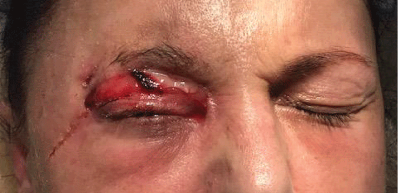
Surgery was performed on December 24, 2022, to drain an abscess in the right eye area. Under local anaesthesia, an abscess was opened on the upper lateral edge of the eye socket. About 5 ml of purulent exudate was released, and the wound was washed with an antiseptic and drained with a rubber tape graduate. Hemostasis during the operation. A bandage with an antiseptic solution is applied (Figure 1).
Figure 1: The general appearance of the face of patient N. after opening the phlegmon of the periorbital area on the right.
Biochemical analysis of blood (24.12.24). AlAT 32.48 U/l, AsAT 54.03 U/l, total bilirubin 10.72 mcmol/l, direct bilirubin 5.42 mcmol/l, blood glucose 6.47 mmol/l, calcium in blood serum 2.4 mmol/l, potassium – 2.8 mmol/l, magnesium 0.71 mmol/l, sodium 128.0 mmol/l, creatinine 46.02 mcmol/l, urea 0.71 mmol/l, cholesterol 5.0 mmol/l.
Microbiological examination of the wound contents confirmed the growth of Staphylococcus epidermidis and GroupA beta-hemolytic Streptococci (GAS).
From 12/26/2022 to 01/06/2023, the patient noted a gradual decrease in pain in the wound area and a decrease in body temperature. Objectively, the amount of purulent secretions from the wound decreased, and the hyperemia of the facial tissues decreased significantly. The area of necrosis was constantly treated with an antiseptic, and the drains were replaced. Aseptic bandages were applied to the wound.
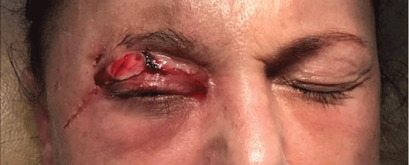
Status localis: (January 6, 2023). Necrotic masses from the right eyelid have moved away on their own; there is no epidermis in the upper eyelid area, and the tissue is granulating (Figure 2).
Figure 2: Appearance of the postoperative wound after 14 days.
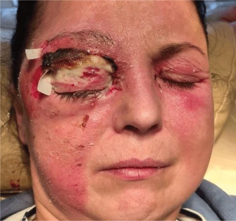
Our experience allows us to state that closing the wound with auto skin after removing necrotic masses can limit the mobility of the upper eyelid in similar situations. This significantly reduces the quality of life and requires additional corrective operations on the upper eyelid to restore mobility. Therefore, on January 7, 2023, we closed the wound on the upper eyelid with an A-PRF membrane prepared according to the standard method (Figure 3).
Figure 3: View of the postoperative wound after its closure with A-PRF membrane.
The postoperative period was smooth and uneventful. The skin of the upper eyelid has completely recovered, and the eyelid's mobility has been preserved. Laboratory indicators of general and biochemical blood analysis were within normal limits.
On January 21, 2023, the patient was discharged in a satisfactory state. An ophthalmologist's observation is recommended. Further monitoring of the wound's healing could not be carried out as the patient left the borders of Ukraine.
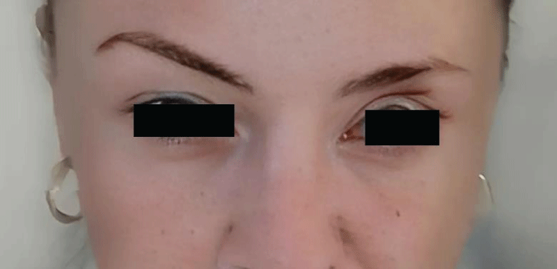
Examination of the patient on April 14, 2023, in remote mode (temporarily living in Germany) confirmed the complete renewal of mobility of the right upper eyelid and preservation of vision. However, there is scarring on the upper left eyelid, which was intact during the patient's stay in the clinic (Figure 4).
Figure 4: The patient's facial appearance 3,5 months post-surgery.
Erysipelas, a bacterial skin infection, is a type of cellulitis that impacts the outermost layers of the skin on various body parts such as the face, legs, arms, and torso [88Michael Y, Shaukat NM. Erysipelas. 2023 Aug 7. In: StatPearls [Internet]. Treasure Island (FL): StatPearls Publishing; 2024 Jan–. PMID: 30335280.]. Erysipelas can affect people of all age groups, races, and sexes. Some studies have shown that erysipelas is more common in females [99Plagens-Rotman K, Przybylska R, Gerke K, Adamski Z, Czarnecka-Operacz M. 55-year old woman with erysipelas. Postepy Dermatol Alergol. 2020 Aug;37(4):613-616. doi: 10.5114/ada.2020.98225. Epub 2020 Sep 2. PMID: 32994788; PMCID: PMC7507150.]. It can affect all age groups but is most common in the extremes of age [1010Krasagakis K, Valachis A, Maniatakis P, Krüger-Krasagakis S, Samonis G, Tosca AD. Analysis of epidemiology, clinical features and management of erysipelas. Int J Dermatol. 2010 Sep;49(9):1012-7. doi: 10.1111/j.1365-4632.2010.04464.x. PMID: 20931671.]. Recurrent erysipelas have also been reported, with the infection typically reoccurring at the same site.
The range of potential diagnosing for erysipelas can be extensive. Erythema, warmth, and oedema can indicate various conditions [1111Scarano A, Inchingolo F, Scogna G, Leo L, Crisante A, Greco Lucchina A, Lorusso F. Xanthelasma palpebrarum removed with Atmospheric Plasma technique: 11-year follow up. J Biol Regul Homeost Agents. 2021 Mar-Apr;35(2 Suppl. 1):181-185. doi: 10.23812/21-2supp1-18. PMID: 34281315.,1212Datta I, Casanas B, Vincent AL, Greene JN. The red face: Erysipelas versus, parvovirus B19, SLE, and rosacea. Asian Biomed. 2009 Dec 1; 3:681-8.]. Some more prevalent conditions include cellulitis, erysipeloid, impetigo, herpes zoster, and necrotizing fasciitis.
In a study by Buckland, Golden T. et al. a 62-year-old woman with rheumatoid arthritis developed chronic lymphedema around the eyes two months after being infected with Group A β-hemolytic streptococcus in the face [1313Buckland GT 3rd, Carlson JA, Meyer DR. Persistent periorbital and facial lymphedema associated with Group A beta-hemolytic streptococcal infection (erysipelas). Ophthalmic Plast Reconstr Surg. 2007 Mar-Apr;23(2):161-3. doi: 10.1097/01.iop.0000256161.79015.38. PMID: 17413641.]. A study by Nadeem A. et al. described a 57-year-old man diagnosed with erysipelas in the Emergency Department [1414Nadeem A, Espinosa JA, Lucerna AA. Case Report: Erysipelas Diagnosed in the Emergency Department.].
The suggested therapy for erysipelas involves antibiotics, particularly semi-synthetic penicillin [1515Brindle R, Williams OM, Barton E, Featherstone P. Assessment of Antibiotic Treatment of Cellulitis and Erysipelas: A Systematic Review and Meta-analysis. JAMA Dermatol. 2019 Sep 1;155(9):1033-1040. doi: 10.1001/jamadermatol.2019.0884. PMID: 31188407; PMCID: PMC6563587.]. The incidence of erysipelas has declined following the introduction of antibiotics and advancements in sanitation [1616Klotz C, Courjon J, Michelangeli C, Demonchy E, Ruimy R, Roger PM. Adherence to antibiotic guidelines for erysipelas or cellulitis is associated with a favorable outcome. Eur J Clin Microbiol Infect Dis. 2019 Apr;38(4):703-709. doi: 10.1007/s10096-019-03490-6. Epub 2019 Jan 26. PMID: 30685804.].
Surgery is typically necessary only in rare cases of erysipelas when the infection has rapidly progressed and led to the death of healthy tissue. In such instances, a surgical procedure may be needed to excise or remove the dead tissue.
The potential of advanced platelet-rich fibrin (A-PRF) in tissue regeneration is significant, offering a hopeful outlook for advanced treatment options. This concentration of autologous platelets on a fibrin membrane, without added external factors, has shown promising results for tissue regeneration [1717Miron RJ, Pinto NR, Quirynen M, Ghanaati S. Standardization of relative centrifugal forces in studies related to platelet-rich fibrin. J Periodontol. 2019 Aug;90(8):817-820. doi: 10.1002/JPER.18-0553. Epub 2019 Mar 1. PMID: 30730050.].
The A-PRF membrane has been successfully utilized in clinical settings for various purposes associated with reconstructive and jaw graft surgeries [1818Clark D, Rajendran Y, Paydar S, Ho S, Cox D, Ryder M, Dollard J, Kao RT. Advanced platelet-rich fibrin and freeze-dried bone allograft for ridge preservation: A randomized controlled clinical trial. J Periodontol. 2018 Apr;89(4):379-387. doi: 10.1002/JPER.17-0466. PMID: 29683498; PMCID: PMC6483085.]. Its high potential to stimulate tissue growth, enriched with leukocytes and cell growth factors, reassures its effectiveness. Moreover, its use of the body's own material eliminates significant economic costs [1919Caruana A, Savina D, Macedo JP, Soares SC. From Platelet-Rich Plasma to Advanced Platelet-Rich Fibrin: Biological Achievements and Clinical Advances in Modern Surgery. Eur J Dent. 2019 May;13(2):280-286. doi: 10.1055/s-0039-1696585. Epub 2019 Sep 11. PMID: 31509878; PMCID: PMC6777161.].
The disease spreading as gangrene or fasciitis with a significant septic syndrome is the most feared systemic complication of erysipelas [2020Titou H, Ebongo C, Bouati E, Boui M. Risk factors associated with local complications of erysipelas: a retrospective study of 152 cases. Pan Afr Med J. 2017 Feb 5;26:66. doi: 10.11604/pamj.2017.26.66.11096. PMID: 28451043; PMCID: PMC5398858.]. These complications can be life-threatening and underscore the importance of early diagnosis and prompt treatment of erysipelas.
The presented case of primary gangrenous-phlegmonous erysipelas of the face with necrosis of the upper eyelid highlights the importance of recognizing atypical manifestations of erysipelas. Clinical and laboratory-instrumental manifestations in this case were atypical, leading to diagnostic challenges. Understanding and identifying such unusual presentations are essential for prompt and effective surgical and therapeutic interventions. This knowledge equips medical professionals with the necessary skills to manage similar cases in the future, emphasizing the significance of continuous medical education and awareness of rare and severe forms of erysipelas. Further research and reporting of similar cases can contribute to a better understanding and management of these challenging clinical scenarios.
The study protocols were approved by the Bioethics Committee of I. Horbachevsky Ternopil National Medical University.
All the patient investigations conformed to the principles outlined in the Council of Europe Convention on Human Rights and Biomedicine (April 4, 1997), the World Health Association Helsinki Declaration on Ethical Principles for Scientific Research with Human Participation (1964-2000), and the Order of the Ministry of Health of Ukraine No. 281 of November 1, 2000; Declaration of Helsinki “World Medical Association Declaration of Helsinki Ethical Principles for Medical Research Involving Human Subjects” (2001), Code of Scientist of Ukraine (2009). All patients’ examinations were done with informed consent.
Patient consent statement
Written patient informed consent was obtained for publication.
Consent to publication
Informed consent to publication was obtained from relevant participants.
Availability of data and materials
The datasets for this study will be available at a reasonable request to the corresponding author.
Hallab L, Taleb B. Facial Erysipelas: A Case Report. Integrative Journal of Medical Sciences. 2021;8. https://doi.org/10.15342/ijms.2021.315
Ezemma O, Korman AM, Wang HE, Kaffenberger B. Diagnostic methods for the confirmation of non-purulent cellulitis: a review. Arch Dermatol Res. 2023 Nov;315(9):2519-2527. doi: 10.1007/s00403-023-02658-7. Epub 2023 Jul 8. PMID: 37421422.
Brishkoska-Boshkovski V, Dimitrovska I, Kondova-Topuzovska I. Clinical Presentation and Laboratory Characteristics in Acute and Recurrent Erysipelas. Open Access Maced J Med Sci. 2019 Mar 14;7(5):771-774. doi: 10.3889/oamjms.2019.213. PMID: 30962836; PMCID: PMC6447339.
Kozłowska D, Myśliwiec H, Kiluk P, Baran A, Milewska AJ, Flisiak I. Clinical and epidemiological assessment of patients hospitalized for primary and recurrent erysipelas. Przegl Epidemiol. 2016;70(4):575-584. English, Polish. PMID: 28221013.
Andreychyn MA(Ed.). Infectious diseases in general practice and family medicine. TDMU "Ukrmedknyha" 2007 [in Ukrainian].
Sadighi Akha AA, Csomós K, Ujházi B, Walter JE, Kumánovics A. Evolving Approach to Clinical Cytometry for Immunodeficiencies and Other Immune Disorders. Clin Lab Med. 2023 Sep;43(3):467-483. doi: 10.1016/j.cll.2023.05.002. Epub 2023 Jun 9. PMID: 37481324.
Shapoval SD, Vasylevska LA, Bielinska VO. Clinical features and principles of differential diagnosis of erysipelas. The Ukrainian Journal of Clinical Surgery. 2021 Mar 28; 88(1-2):61-8. doi: 10.26779/2522-1396.2021.1-2.61
Michael Y, Shaukat NM. Erysipelas. 2023 Aug 7. In: StatPearls [Internet]. Treasure Island (FL): StatPearls Publishing; 2024 Jan–. PMID: 30335280.
Plagens-Rotman K, Przybylska R, Gerke K, Adamski Z, Czarnecka-Operacz M. 55-year old woman with erysipelas. Postepy Dermatol Alergol. 2020 Aug;37(4):613-616. doi: 10.5114/ada.2020.98225. Epub 2020 Sep 2. PMID: 32994788; PMCID: PMC7507150.
Krasagakis K, Valachis A, Maniatakis P, Krüger-Krasagakis S, Samonis G, Tosca AD. Analysis of epidemiology, clinical features and management of erysipelas. Int J Dermatol. 2010 Sep;49(9):1012-7. doi: 10.1111/j.1365-4632.2010.04464.x. PMID: 20931671.
Scarano A, Inchingolo F, Scogna G, Leo L, Crisante A, Greco Lucchina A, Lorusso F. Xanthelasma palpebrarum removed with Atmospheric Plasma technique: 11-year follow up. J Biol Regul Homeost Agents. 2021 Mar-Apr;35(2 Suppl. 1):181-185. doi: 10.23812/21-2supp1-18. PMID: 34281315.
Datta I, Casanas B, Vincent AL, Greene JN. The red face: Erysipelas versus, parvovirus B19, SLE, and rosacea. Asian Biomed. 2009 Dec 1; 3:681-8.
Buckland GT 3rd, Carlson JA, Meyer DR. Persistent periorbital and facial lymphedema associated with Group A beta-hemolytic streptococcal infection (erysipelas). Ophthalmic Plast Reconstr Surg. 2007 Mar-Apr;23(2):161-3. doi: 10.1097/01.iop.0000256161.79015.38. PMID: 17413641.
Nadeem A, Espinosa JA, Lucerna AA. Case Report: Erysipelas Diagnosed in the Emergency Department.
Brindle R, Williams OM, Barton E, Featherstone P. Assessment of Antibiotic Treatment of Cellulitis and Erysipelas: A Systematic Review and Meta-analysis. JAMA Dermatol. 2019 Sep 1;155(9):1033-1040. doi: 10.1001/jamadermatol.2019.0884. PMID: 31188407; PMCID: PMC6563587.
Klotz C, Courjon J, Michelangeli C, Demonchy E, Ruimy R, Roger PM. Adherence to antibiotic guidelines for erysipelas or cellulitis is associated with a favorable outcome. Eur J Clin Microbiol Infect Dis. 2019 Apr;38(4):703-709. doi: 10.1007/s10096-019-03490-6. Epub 2019 Jan 26. PMID: 30685804.
Miron RJ, Pinto NR, Quirynen M, Ghanaati S. Standardization of relative centrifugal forces in studies related to platelet-rich fibrin. J Periodontol. 2019 Aug;90(8):817-820. doi: 10.1002/JPER.18-0553. Epub 2019 Mar 1. PMID: 30730050.
Clark D, Rajendran Y, Paydar S, Ho S, Cox D, Ryder M, Dollard J, Kao RT. Advanced platelet-rich fibrin and freeze-dried bone allograft for ridge preservation: A randomized controlled clinical trial. J Periodontol. 2018 Apr;89(4):379-387. doi: 10.1002/JPER.17-0466. PMID: 29683498; PMCID: PMC6483085.
Caruana A, Savina D, Macedo JP, Soares SC. From Platelet-Rich Plasma to Advanced Platelet-Rich Fibrin: Biological Achievements and Clinical Advances in Modern Surgery. Eur J Dent. 2019 May;13(2):280-286. doi: 10.1055/s-0039-1696585. Epub 2019 Sep 11. PMID: 31509878; PMCID: PMC6777161.
Titou H, Ebongo C, Bouati E, Boui M. Risk factors associated with local complications of erysipelas: a retrospective study of 152 cases. Pan Afr Med J. 2017 Feb 5;26:66. doi: 10.11604/pamj.2017.26.66.11096. PMID: 28451043; PMCID: PMC5398858.
Levkiv M, Nahirnyi Y, Kopcha V, Tverdokhlib N, Stefaniv I. A Case of Facial Erysipelas with Necrosis of the Upper Eyelid. IgMin Res. . 02 Sep, 2024; 2(9): 739-743. IgMin ID: igmin241; DOI:10.61927/igmin241; Available at: igmin.link/p241
1Dental Surgery Department, I Horbachevsky Ternopil National Medical University, Ternopil 46003, Ukraine
2Department of Infectious Diseases with Epidemiology, Skin and Venereal Diseases, I Horbachevsky Ternopil National Medical University, Ternopil 46001, Ukraine
3Dental Therapy Department, I Horbachevsky Ternopil National Medical University, Ternopil 46003, Ukraine
Address Correspondence: Mariana Levkiv, Dental Therapy Department, I Horbachevsky Ternopil National Medical University, Ternopil 46003, Ukraine, Email: [email protected]
How to cite this article: Levkiv M, Nahirnyi Y, Kopcha V, Tverdokhlib N, Stefaniv I. A Case of Facial Erysipelas with Necrosis of the Upper Eyelid. IgMin Res. . 02 Sep, 2024; 2(9): 739-743. IgMin ID: igmin241; DOI:10.61927/igmin241; Available at: igmin.link/p241
Figure 1: The general appearance of the face of patient N. a...
Figure 2: Appearance of the postoperative wound after 14 day...
Figure 3: View of the postoperative wound after its closure ...
Figure 4: The patient's facial appearance 3,5 months post-su...
Hallab L, Taleb B. Facial Erysipelas: A Case Report. Integrative Journal of Medical Sciences. 2021;8. https://doi.org/10.15342/ijms.2021.315
Ezemma O, Korman AM, Wang HE, Kaffenberger B. Diagnostic methods for the confirmation of non-purulent cellulitis: a review. Arch Dermatol Res. 2023 Nov;315(9):2519-2527. doi: 10.1007/s00403-023-02658-7. Epub 2023 Jul 8. PMID: 37421422.
Brishkoska-Boshkovski V, Dimitrovska I, Kondova-Topuzovska I. Clinical Presentation and Laboratory Characteristics in Acute and Recurrent Erysipelas. Open Access Maced J Med Sci. 2019 Mar 14;7(5):771-774. doi: 10.3889/oamjms.2019.213. PMID: 30962836; PMCID: PMC6447339.
Kozłowska D, Myśliwiec H, Kiluk P, Baran A, Milewska AJ, Flisiak I. Clinical and epidemiological assessment of patients hospitalized for primary and recurrent erysipelas. Przegl Epidemiol. 2016;70(4):575-584. English, Polish. PMID: 28221013.
Andreychyn MA(Ed.). Infectious diseases in general practice and family medicine. TDMU "Ukrmedknyha" 2007 [in Ukrainian].
Sadighi Akha AA, Csomós K, Ujházi B, Walter JE, Kumánovics A. Evolving Approach to Clinical Cytometry for Immunodeficiencies and Other Immune Disorders. Clin Lab Med. 2023 Sep;43(3):467-483. doi: 10.1016/j.cll.2023.05.002. Epub 2023 Jun 9. PMID: 37481324.
Shapoval SD, Vasylevska LA, Bielinska VO. Clinical features and principles of differential diagnosis of erysipelas. The Ukrainian Journal of Clinical Surgery. 2021 Mar 28; 88(1-2):61-8. doi: 10.26779/2522-1396.2021.1-2.61
Michael Y, Shaukat NM. Erysipelas. 2023 Aug 7. In: StatPearls [Internet]. Treasure Island (FL): StatPearls Publishing; 2024 Jan–. PMID: 30335280.
Plagens-Rotman K, Przybylska R, Gerke K, Adamski Z, Czarnecka-Operacz M. 55-year old woman with erysipelas. Postepy Dermatol Alergol. 2020 Aug;37(4):613-616. doi: 10.5114/ada.2020.98225. Epub 2020 Sep 2. PMID: 32994788; PMCID: PMC7507150.
Krasagakis K, Valachis A, Maniatakis P, Krüger-Krasagakis S, Samonis G, Tosca AD. Analysis of epidemiology, clinical features and management of erysipelas. Int J Dermatol. 2010 Sep;49(9):1012-7. doi: 10.1111/j.1365-4632.2010.04464.x. PMID: 20931671.
Scarano A, Inchingolo F, Scogna G, Leo L, Crisante A, Greco Lucchina A, Lorusso F. Xanthelasma palpebrarum removed with Atmospheric Plasma technique: 11-year follow up. J Biol Regul Homeost Agents. 2021 Mar-Apr;35(2 Suppl. 1):181-185. doi: 10.23812/21-2supp1-18. PMID: 34281315.
Datta I, Casanas B, Vincent AL, Greene JN. The red face: Erysipelas versus, parvovirus B19, SLE, and rosacea. Asian Biomed. 2009 Dec 1; 3:681-8.
Buckland GT 3rd, Carlson JA, Meyer DR. Persistent periorbital and facial lymphedema associated with Group A beta-hemolytic streptococcal infection (erysipelas). Ophthalmic Plast Reconstr Surg. 2007 Mar-Apr;23(2):161-3. doi: 10.1097/01.iop.0000256161.79015.38. PMID: 17413641.
Nadeem A, Espinosa JA, Lucerna AA. Case Report: Erysipelas Diagnosed in the Emergency Department.
Brindle R, Williams OM, Barton E, Featherstone P. Assessment of Antibiotic Treatment of Cellulitis and Erysipelas: A Systematic Review and Meta-analysis. JAMA Dermatol. 2019 Sep 1;155(9):1033-1040. doi: 10.1001/jamadermatol.2019.0884. PMID: 31188407; PMCID: PMC6563587.
Klotz C, Courjon J, Michelangeli C, Demonchy E, Ruimy R, Roger PM. Adherence to antibiotic guidelines for erysipelas or cellulitis is associated with a favorable outcome. Eur J Clin Microbiol Infect Dis. 2019 Apr;38(4):703-709. doi: 10.1007/s10096-019-03490-6. Epub 2019 Jan 26. PMID: 30685804.
Miron RJ, Pinto NR, Quirynen M, Ghanaati S. Standardization of relative centrifugal forces in studies related to platelet-rich fibrin. J Periodontol. 2019 Aug;90(8):817-820. doi: 10.1002/JPER.18-0553. Epub 2019 Mar 1. PMID: 30730050.
Clark D, Rajendran Y, Paydar S, Ho S, Cox D, Ryder M, Dollard J, Kao RT. Advanced platelet-rich fibrin and freeze-dried bone allograft for ridge preservation: A randomized controlled clinical trial. J Periodontol. 2018 Apr;89(4):379-387. doi: 10.1002/JPER.17-0466. PMID: 29683498; PMCID: PMC6483085.
Caruana A, Savina D, Macedo JP, Soares SC. From Platelet-Rich Plasma to Advanced Platelet-Rich Fibrin: Biological Achievements and Clinical Advances in Modern Surgery. Eur J Dent. 2019 May;13(2):280-286. doi: 10.1055/s-0039-1696585. Epub 2019 Sep 11. PMID: 31509878; PMCID: PMC6777161.
Titou H, Ebongo C, Bouati E, Boui M. Risk factors associated with local complications of erysipelas: a retrospective study of 152 cases. Pan Afr Med J. 2017 Feb 5;26:66. doi: 10.11604/pamj.2017.26.66.11096. PMID: 28451043; PMCID: PMC5398858.